Tendon Diagram Of Wrist - Pin on Anatomical references. Wrist tendonitis is the inflammation of a tendon in the wrist. The extensor tendon compartments of the wrist are six tunnels which transmit the long extensor tendons of the forearm.they are located on the posterior aspect of the wrist. 297 x 320 jpeg 25 кб. Consider physical therapy for wrist tendonitis for an individualized exercise program if your pain persists or if your symptoms are accompanied by numbness or tingling. Extensor tendon compartments of the wrist are anatomical tunnels on the back of the wrist that contain tendons of muscles that extend (as opposed to flex) the wrist and the digits (fingers and thumb).
There are several tendons that surround the wrist joint that can become injured or inflamed. Tendons are thick, fibrous cords that connect muscle to bone. How to tell if my wrist tendon is torn and what level it is? answered by dr. Or if it would even be considered a sprain still. Consider physical therapy for wrist tendonitis for an individualized exercise program if your pain persists or if your symptoms are accompanied by numbness or tingling.
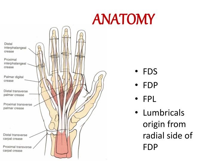
PPT - Cut Wrist & Flexor tendon injury PowerPoint Presentation - ID:7081690 from image3.slideserve.com Notably displays the transverse carpal ligament (flexor retinaculum) and the palmar fascia. Tendon diagrams and design force vectors. Pinky side of the wrist. This is a tenosynovitis affecting the tendons on the thumb side of the wrist. The wrist tendons slide through smooth sheaths as they pass by the wrist joint. This tendon is one of two tendons that bend the wrist. How to tell if my wrist tendon is torn and what level it is? answered by dr. Tendons are tissues that attach our muscles to our bones.
Each tunnel is lined internally by a synovial sheath and separated from one another by a fibrous septa.
Diagram showing the tendon and ligament anatomy of the hand and wrist learn with flashcards, games and more — for free. If any wrist tendonitis exercises cause you pain, stop immediately. Tendons are thick, fibrous cords that connect muscle to bone. This tendon is one of two tendons that bend the wrist. (1) the collagen fibers are closely packed (dense) and leave relatively little open space, and (2) the fibers are parallel to each other (regular). Diagrammatic representation of the wrist extensor compartments shows the spatial relationship of the six extensor compartments. 3 schematic representation of the hierarchical. Use the mouse scroll wheel to move the images up and down alternatively use the tiny arrows (>>) on both side of the image to move the images. How to tell if my wrist tendon is torn and what level it is? answered by dr. Wrist tendonitis is the inflammation of one or more tendons in the wrist. Wrist tendonitis is not necessarily confined to a single tendon or part of the wrist. Tendons are fibrous cords attached to muscles and bone. Tendons are thick, fibrous cords that connect muscles to bones.
Trauma to the tendon or inflammation can cause the sheath to stiffen and swell. Wrist tendonitis is not necessarily confined to a single tendon or part of the wrist. Tendons are thick, fibrous cords that connect muscles to bones. I felt something shift a bit, no swelling whatsoever. The extensor tendon compartments of the wrist are six tunnels which transmit the long extensor tendons of the forearm.they are located on the posterior aspect of the wrist.
Schematic diagram of volar extrinsic wrist ligaments. RSC volar... | Download Scientific Diagram from www.researchgate.net Wrist tendonitis is not necessarily confined to a single tendon or part of the wrist. … this diagram with labels depicts and explains the. Posterior calf anatomy muscles of the lower leg diagram. Perform wrist exercises after the initial pain has subsided. Anatomy atlas of the upper limb: Ankle tendon anatomy, elbow tendon anatomy, forearm tendon anatomy, wrist flexor tendon anatomy, wrist tendon anatomy mri, wrist tendon anatomy pictures, wrist tendon pain, wrist tendonitis, hand, ankle tendon anatomy related posts of wrist tendon anatomy diagrams. Tendons in the wrist work by sliding through tendon sheaths that are covered with synovial fluid. Tendon diagrams and design force vectors.
They can become swollen and sore from over use.
Tendon diagrams and design force vectors. The tendons that control movement in your hands, wrists and fingers run through your forearm. Related pictures anterior and posterior. Tendons are thick, fibrous cords that connect muscle to bone. Extensor tendon compartments of the wrist are anatomical tunnels on the back of the wrist that contain tendons of muscles that extend (as opposed to flex) the wrist and the digits (fingers and thumb). (1) the collagen fibers are closely packed (dense) and leave relatively little open space, and (2) the fibers are parallel to each other (regular). This tendon is one of two tendons that bend the wrist. When muscles contract, they pull on the tendons to. Long flexor tendons extend from the forearm muscles through the wrist and attach to the small bones of the fingers and thumb. These tendon sheaths allow the tendons to glide smoothly as the diagnosis of wrist tendonitis is made by looking for the characteristic signs of this condition. The parallel arrangement of fibers is an adaptation to the fact that. Epidemiology there is a 3:1 male predominance and arise between the 2nd to 5th decades 2. 34 834 просмотра 34 тыс.
In addition, depending on the tendon that is inflamed, the. Tendons are tissues that attach our muscles to our bones. There are several tendons that surround the wrist joint that can become injured or inflamed. This tendon is one of two tendons that bend the wrist. Anatomy diagrams of shoulder, arm, elbow, forearm, wrist and hand.
Arterial circulation in the forearm and hand. | Download Scientific Diagram from www.researchgate.net The extensor tendon compartments of the wrist are six tunnels which transmit the long extensor tendons of the forearm.they are located on the posterior aspect of the wrist. The parallel arrangement of fibers is an adaptation to the fact that. It attaches to the base of the second and third hand bones. This mri wrist coronal cross sectional anatomy tool is absolutely free to use. 297 x 320 jpeg 25 кб. This tendon works with the ecrb and ecrl to straighten the wrist. Tendonitis usually develops as a result of acute or repetitive injury to a tendon. This is a tenosynovitis affecting the tendons on the thumb side of the wrist.
Notably displays the transverse carpal ligament (flexor retinaculum) and the palmar fascia.
(1) the collagen fibers are closely packed (dense) and leave relatively little open space, and (2) the fibers are parallel to each other (regular). The biceps human anatomy function tendons of the posterior dorsal wrist medical images for. Each tunnel is lined internally by a synovial sheath and separated from one another by a fibrous septa. Tendons are fibrous cords attached to muscles and bone. Related pictures anterior and posterior. Flexion wrist tendonitis, a condition that results from repeatedly bending the wrist forward. This tendon is one of two tendons that bend the wrist. The tendons that control movement in your hands, wrists and fingers run through your forearm. The wrist tendons slide through smooth sheaths as they pass by the wrist joint. These tendon sheaths allow the tendons to glide smoothly as the diagnosis of wrist tendonitis is made by looking for the characteristic signs of this condition. From the momentum my wrist rolled over and turned over my brake. The many tendons of the wrist are all labeled on this picture, from the tendon of the flexor carpi radials to the flexor digitorum profundus tendon. Tendons in the wrist work by sliding through tendon sheaths that are covered with synovial fluid.
When muscles contract, they pull on the tendons to tendon diagram. This tendon is one of two tendons that bend the wrist.
Share :
Post a Comment
for "Tendon Diagram Of Wrist - Pin on Anatomical references"


Post a Comment for "Tendon Diagram Of Wrist - Pin on Anatomical references"